A honey bee colony does not follow a calendar. Should its model?

New BEEHAVE-PPE links egg-laying rates to pollen foraging, brood size, and pheromone feedback, not just average weather.

Guest blog post by Dominik Lammers

Models help us ask a deceptively simple question: if the world works in a particular way, what should we expect to happen?

For honey bee colonies, answering that question is difficult. A colony’s development depends on food, weather, disease, the surrounding landscape, and the behaviour of thousands of individual bees. These influences do not act separately. They affect one another, sometimes in ways that are hard to isolate even in carefully designed experiments.

In a recent study with Fabrice Requier, Andreas Focks, and Jürgen Groeneveld, we explored one of these connections using BEEHAVE, a computer model of a honey bee colony. We developed an exploratory extension called BEEHAVE-PPE, asking whether seasonal colony development could arise from links between pollen, brood pheromones, egg laying, and temperature, rather than following a seasonal egg-laying pattern specified in advance.

The result is not a finished replacement for the original model. It is a hypothesis about processes inside the hive: one that can produce plausible colony dynamics, while also making clear where scientific understanding remains incomplete.

A starting point that made BEEHAVE useful

Wooden beehive boxes for beekeeping and honey collecting in blooming canola field.
Wooden beehive boxes for beekeeping and honey collecting in blooming canola field. Credit to stevanovicigor via Envato.

BEEHAVE has been used to investigate how food availability, weather, parasites, pesticides, and beekeeping practices can affect honey bee colonies. It links processes inside the hive with conditions in the surrounding landscape, allowing researchers to explore combinations of stressors that would be difficult to study directly in real colonies.

To give a simulated colony a realistic seasonal trajectory, the original BEEHAVE uses an annual curve that determines how many eggs the queen lays on each day of the year. This was an effective modelling choice. It allowed the colony to develop in a broadly realistic way under typical Central European conditions and made it possible to investigate many other questions.

At the same time, this relationship shapes much of the simulated colony’s development. The queen follows a known seasonal pattern, while real colonies are likely to adjust reproduction in response to conditions inside and outside the hive.

I was interested in what would happen if that central pattern was no longer specified in advance. That became less like removing a single line from a model and more like beginning a journey. Each change exposed a new problem: without the fixed curve, what would initiate colony growth? What would prevent it from continuing indefinitely? What processes might connect the colony’s nutritional state to its reproduction?

For me, this was both a creative and an intellectual task. It involved imagining possible biological explanations, searching the literature for evidence that could support them, and translating those ideas into code. BEEHAVE-PPE emerged from that process.

A feedback loop inside the hive

The resulting model is built around three linked ideas.

Conceptual overview of the relationships between stored pollen and egg-laying in BEEHAVE and the new version, BEEHAVE-PPE. Major differences are indicated in red. Credit to Lammers et al., 2026.

First, the amount of pollen stored in the colony influences the queen’s egg laying. Pollen is the main protein source for feeding brood, so a colony with more available pollen can plausibly support more reproduction.

Second, larvae produce brood pheromones: chemical signals that can influence the behaviour of worker bees.

Third, brood pheromones can encourage workers to collect more pollen rather than nectar. This helps replenish pollen stores and can support further egg laying.

Together, these processes create a feedback loop:

pollen availability -> egg laying -> brood pheromones -> pollen collection -> pollen availability

Temperature affects this loop. In BEEHAVE-PPE, warmer conditions increase the assumed degradation of brood pheromone. This weakens the signal encouraging pollen collection and can slow colony growth.

In one sense, temperature fulfills a role similar to the original seasonal egg-laying curve: it helps shape when growth slows and when a colony reaches its annual peak. But it does so differently. Rather than instructing the queen to lay a certain number of eggs on a particular date, it represents an environmental condition that can differ between places and years. This opens the possibility that the same underlying model could respond differently under different temperature regimes, provided that its temperature relationships can eventually be tested and calibrated.

Letting seasonal dynamics emerge

Bees Entering Hive
Bees Entering Hive on Wooden Frame. Credit to NaturesCharm via Envato.

Under the average weather conditions used in this study, BEEHAVE-PPE produced plausible seasonal colony dynamics. The simulated colonies grew in spring, reached a population peak in early summer, and declined afterwards. The number of adult bees in the simulated colonies followed the broad timing and shape of the French monitoring data used to calibrate the new module.

The model did not prove that real colonies work through precisely this mechanism. It cannot do that. A model can show that a proposed explanation is capable of generating an observed pattern; it cannot establish, by itself, that the explanation is the only or exact one used in nature.

What BEEHAVE-PPE does show is that the seasonal development of a colony need not be prescribed as a curve from the outset. A plausible combination of pollen availability, brood signalling, worker behaviour, and temperature can generate it.

That changes the role of the model. Instead of only reproducing a known seasonal pattern, it asks what biological connections could be responsible for that pattern.

Learning from where the model fails

bees on honeycomb
Bees on honeycomb. Credit to Kohanova via Envato.

The model also revealed a clear limitation. It produced plausible dynamics under averaged weather conditions, but it was vulnerable to prolonged periods in which bees could not collect pollen.

In the simulation, a long interruption weakens the feedback loop. Less pollen leads to reduced egg laying; fewer larvae produce less brood pheromone; and the weaker pheromone signal reduces the incentive to collect pollen when conditions improve. The colony can become trapped in a low-pollen, low-brood state.

This is unlikely to be the complete story in real colonies. Colonies can buffer difficult periods through stored resources and changes in brood and worker management. BEEHAVE already contains potentially relevant processes, including brood cannibalism and worker self-metabolism, but these are not yet represented in sufficient nutritional detail to support recovery within the new feedback loop.

That result gives the next steps a clearer direction. Rather than simply knowing that the model behaves unrealistically after sustained poor weather, we can identify the chain of events that causes it. This points to promising improvements, including better representation of nutrient reserves, resource recovery through brood cannibalism, and the colony processes that help it restart reproduction after a difficult period.

The data needed to go further

Honey bees flying into wooden beehive.
Honey bees flying into wooden beehive. Credit to cookelma via Envato.

Developing and testing a model depends on data. The French dataset used in this study, covering 250 colonies, was especially valuable because it provided an unusually broad picture of seasonal colony development under comparable climatic conditions. It made it possible to see not just how one colony behaved, but what a larger set of colonies broadly did over a season.

This kind of baseline information is more limited than it may seem. For several important aspects of honey bee colony development, some of the most detailed observations still trace back to research from decades ago. We have valuable knowledge about individual processes, but less information showing how pollen availability, egg laying, brood development, worker behaviour, temperature, and population size change together over time.

New long-term observation approaches, including continuously monitored colonies, could help fill this gap. Targeted measurements of these linked processes would make it possible to test not only whether BEEHAVE-PPE produces realistic patterns, but whether it does so for the right biological reasons.

Why this matters for future stressor research

BEEHAVE is often used to explore how poor forage, adverse weather, parasites, pesticides, and beekeeping practices may affect colony development. In the original model, these pressures act on a colony whose broad reproductive trajectory is already set by the annual egg-laying pattern.

Honey Bee on flower
Honey Bee on flower. Credit to IciakPhotos via Envato.

BEEHAVE-PPE changes that relationship. Because egg laying, brood production, pollen collection, and colony strength can influence one another, a stressor can affect more than one isolated part of the model. A shortage of pollen, for example, may not only reduce food available on a particular day. It may also reduce reproduction, alter brood signals, change later foraging behaviour, and affect the colony’s capacity to recover.

The current version may respond too strongly when the feedback loop is interrupted. But if these dynamics can be stabilised and supported by stronger empirical evidence, future versions could give a more complete picture of how stressors interact. They could help investigate when the effects of a stressor are amplified by the colony’s internal state, which combinations are most damaging, and where a colony’s natural buffering mechanisms provide protection.

This is particularly relevant as weather patterns, flowering times, forage availability, and temperature change together. BEEHAVE-PPE is not yet a forecasting tool for climate change or colony risk. It is a first step from an imposed seasonal pattern toward interacting biological mechanisms.

Its main value is that it makes both a plausible explanation and its remaining gaps visible. That is one of the strengths of models: they do not only tell us what we can predict. They show us what we still need to understand before prediction becomes possible.

Original source: 

Lammers D, Requier F, Focks A, Groeneveld J (2026) Food for thought: could the queen’s egg-laying rate in the BEEHAVE honey bee model emerge from the effects of brood pheromones, weather conditions, and pollen availability? Individual-based Ecology 2: e185721. https://doi.org/10.3897/ibe.2.185721

How Citizen Science and Fieldwork Rediscovered Papilio alexanor in Southern Italy

The rediscovery of Papilio alexanor in southern Italy marks a historic moment for Italian lepidopterology.

Guest blog post by Paolo Mazzei

For decades, butterfly enthusiasts and lepidopterists across Italy have wondered: could the lost southern populations of the Southern Swallowtail still be out there?

Historical records of Papilio alexanor in Southern Italy were sparse and dated, often dismissed as stray individuals blown in by strong winds from Greece or the Balkans. But for many lepidopterists, alexanor represents a true holy grail – a majestic, bright-yellow species that is strictly protected.

Our research didn’t start in a lab, but with a smartphone screen. 

When Citizen Science Meets Expert Curiosity

male Papilio alexanor
Male Papilio alexanor on C. ruber. Picture by Stefano Meraglia.

In May 2023, a marine biologist kayaking along the rugged cliffs of Basilicata uploaded a brief video to iNaturalist. It captured an unmistakable female Papilio alexanor fluttering around a giant fennel (Ferula) stalk.

A year later, a second record appeared on iNaturalist: a pristine, freshly emerged female photographed by a German tourist in Campania. Together, these two sightings hinted that the species might be more than a mere vagrant here.

Papilio alexanor
Papilio alexanor. Picture by Paolo Mazzei.

This is where the power of modern biodiversity platforms like iNaturalist, Observation.org, and the eBMS (European Butterfly Monitoring Scheme) shines: they act as distributed networks of eyes on the ground, catching subtle shifts in nature that a single research team could easily miss. Inspired by these community sightings, our team from the Italian Lepidopterological Association (ALI), in collaboration with the University of Turin, decided it was time to step in.

A 1,400 km Journey into the Wild

Obtaining research permits for a strictly protected species under EU law takes time, ethics, and rigor. With support from the University of Turin, we secured formal authorization from the Italian Ministry of Environment in early 2026.

Driven by passion, our team funded the expedition ourselves, driving over 1,400 km round trip to systematically survey the steep coastal cliffs and dry habitats along the Campania–Basilicata border.

  • Pupa of Papilio alexanor.
  • Papilio alexanor caterpillar
  • Papilio alexanor caterpillars feeding on a plant
  • Papilio alexanor caterpillars of different instars, feeding on seeds of a plant
  • Papilio alexanor on a plant

The thrill of discovery was immediate. Standing at a cliffside turnout, we watched the first bright-yellow male soar up from the ravine. Soon we spotted courting pairs feeding on red valerian (Centranthus ruber) and females in the act of ovipositing.

Encouraged, we started scouting similar spots nearby, and the hunch paid off. About 15 to 20 km away, we found a second active breeding colony

Surprising Ecology: A New Host Plant for Italy

Female Papilio alexanor laying eggs on the flowers of F. glauca. Picture by Luciano Valeri.

Our fieldwork revealed two ecological twists, since published in our paper in Nota Lepidopterologica.

First, the Northern Italian populations rely on host plants such as Ptychotis saxifraga or Opopanax chironium, but the southern caterpillars feed exclusively on Ferula glauca. This host-plant association was previously documented in Greece and the Balkans, but never before in Italy.

Second, the flight begins significantly earlier here than in northern populations, reflecting local adaptation to the semi-arid, thermo-Mediterranean climate.

Protecting the Future: Simple Actions for Big Impact

Finding a “lost” population is cause for celebration, but it also brings responsibility. These isolated coastal colonies remain vulnerable to habitat fragmentation, fire, climate extremes, and road infrastructure development.

Female Papilio  alexanor
Female Papilio alexanor on C. ruber. Picture by Andrea Baruzzi, 2026,

To help Papilio alexanor keep thriving here, effective conservation management should prioritise targeted monitoring, integrating structured transects through schemes like eBMS to track population trends over time; habitat protection, preserving coastal slope vegetation and preventing over-mowing or destruction of Ferula glauca stands during roadside maintenance; and responsible citizen engagement, encouraging nature lovers, hikers, and photographers to submit observations with precise geolocation, while ensuring that delicate breeding sites remain respected and undisturbed. 

This story is a reminder that remarkable biological discoveries are still waiting to be made right under our noses. All it takes is a curious eye, a smartphone upload, and a community of passionate researchers ready to follow the trail.

Original source:

Meraglia S, Baruzzi A, Mazzei P, Valeri L, Zerunian Z (2026) First documented record of a reproducing population of Papilio alexanor Esper, 1800 (Lepidoptera, Papilionidae) in Southern Italy, with ecological notes. Nota Lepidopterologica 49: 157-170. https://doi.org/10.3897/nl.49.206084 

Call to contribute to a Special Collection “Vegetation of the Balkan Peninsula” in Vegetation Classification and Survey

A newly launched Special Collection “Vegetation of the Balkan Peninsula” in the open-access, peer-reviewed IAVS journal is now welcoming submissions.

Guest blog post by Jürgen Dengler, Erwin Bergmeier and Urban Šilc

We are pleased to announce the Special Collection on the vegetation of the Balkan Peninsula in IAVS’ gold open access journal Vegetation Classification and Survey (VCS).

This Special Collection is launched in conjunction with the annual conferences of two IAVS Working Groups in 2026, namely the 21st Eurasian Grassland Conference of the EDGG in Sofia, Bulgaria, and the 34th Conference of the European Vegetation Survey (EVS), in Clermont-Ferrand, France.

Within Europe, the vegetation of the Balkan Peninsula is particularly diverse yet understudied in terms of composition, variation and range. With this Special Collection, we thus want to advance the knowledge on the Balkan vegetation, take a fresh look both within and beyond national borders and contribute to an improvement of the “EuroVegChecklist” as the common syntaxonomic backbone of the continent. Following VCS’s scope, contributions either need to develop or to apply a vegetation classification framework of any type. For example, this can be based on a phytosociological classification system, habitat types, or synusiae. Studies on any kind of vegetation from the coasts to the highest mountains, from natural through semi-natural to anthropogenic are welcome.

We envisage particularly the following types of contributions:

  • Revisions of syntaxa (classes, orders, alliances…) at the national or supranational level
  • Vegetation monographs of a certain region
  • Synthetic overviews (category Review and Synthesis) of all syntaxa of a country or other larger regions
  • Nomenclatural proposals related to Balkan syntaxa
  • Habitat typologies and their application
  • Long and Short Database Reports
  • Perspectives on how to harmonize and how to utilize syntaxonomic knowledge

Apart from Research Papers, also Reviews and Syntheses and Forum Papers as well as Long Database Reports and Nomenclatural Proposals are welcome. We hope to see submissions from the Balkan countries (Slovenia, Croatia, Bosnia and Herzegovina, Serbia, Montenegro, Kosovo, North Macedonia, Albania, Bulgaria, Greece and Turkey-in-Europe), while syntheses across multiple countries are particularly welcome. Please consult the author guidelines whose formal requirements also apply to the Special Collection.

Editors: Jürgen Dengler (Switzerland), Erwin Bergmeier (Germany) and Urban Šilc (Slovenia); in case of a high number of submissions, one or two additional guest editors might be appointed.

Procedure and deadlines:

  • Until 31 October 2026: Please submit your (preliminary) abstract online at: https://forms.gle/gxofv8FuZQkqFmeK7. The abstract must follow the VCS Author Guidelines.
  • Until 30 November 2026: The Guest Editors will evaluate the abstracts and inform the authors which manuscripts are invited for submission.
  • Until 30 May 2027: Invited manuscripts can be submitted at: https://vcs.pensoft.net/ 
  • Spring 2028: The Special Collection will be completed with a synthesizing editorial by the Guest Editor team.

Please note that VCS is a gold open access journal where authors are normally required to pay article processing charges (APCs). First authors who are IAVS members receive 50% or 75% discount on regular APCs. If you are an IAVS member but cannot afford the reduced APCs (e.g. because your university does not provide APC funds), you can – after invitation and prior to submission – apply to IAVS’ Global Sponsorship Committee (GSC) for a complete waiver of the APCs, i.e. publishing free of charge. If interested, please get into contact with the chair of the Guest Editors.

We look forward to receiving your contributions and hope that you will join us in making this Special Collection a valuable resource for the IAVS community.

Follow Vegetation Classification and Survey on Facebook and Bluesky to stay up to date on the journal’s latest developments. Or subscribe to the journal’s newsletter via the homepage.

A New Checklist For The Birds Of Calabria: 363 Species At Mediterranean Crossroads

Calabria’s updated bird checklist now lists 363 species, 43 more than in 1993, including Italy’s first documented breeding White-rumped Swift and continued Egyptian Vulture nesting.

Guest blog post by Stazione Ornitologica Calabrese (StOrCal)

Calabria, the toe of the Italian peninsula, lies between the Tyrrhenian and Ionian seas, a short crossing from Sicily and the North African coast. That position has long made it a place where birds from very different regions turn up. Yet its avifauna had not been reviewed in full for more than thirty years.

Our study in Biodiversity Data Journal brings that picture up to date. Drawing on published papers, technical reports, ringing bulletins, museum specimens and validated citizen-science records up to 31 December 2025, we compiled an updated checklist of 363 species and nine subspecies.

That is 43 species more than the previous regional list of 320 (Scebba et al. 1993). None of the older records was dropped, so the additions represent a real gain in regional knowledge.

White-rumped Swift Apus caffer
White-rumped Swift, whose first Italian and central-Mediterranean breeding was documented in Calabria (Pucci et al. 2022). Photo credit: Mario Pucci.

One addition stands out. At an inland site in the province of Crotone, White-rumped Swift (Apus caffer) was found nesting, the first documented breeding of the species in Italy and in the central Mediterranean Basin (Pucci et al. 2022). An African swift breeding this far north suggests Calabria may be a bridgehead for Afro-Iberian species reaching Europe.

Bird ringing
Bird ringing at Punta Alice, where several of the region’s rare vagrants were first detected. Photo credit: Mario Pucci

Most of the other new entries tell a quieter story. Thirty-five of the 43 additions are accidental or vagrant birds, and they point to decades of more systematic fieldwork rather than to genuine range change. Targeted ringing at coastal stations such as Punta Alice (Crotone) and San Michele di Cetraro (Cosenza) has turned up Siberian warblers and flycatchers, among them Pallas’s Leaf Warbler, Dusky Warbler and Red-flanked Bluetail, that would otherwise pass unnoticed. In all, the vagrants recorded in Calabria come from Asia, Africa, northern Europe and even North America, a spread that makes the region a meeting point of separate migration systems.

The checklist also shows what is at stake for conservation. Of the 363 species, 125 are listed in Annex I of the EU Birds Directive, and national Red List categories are available for 230, including nine Critically Endangered.

Egyptian Vulture Neophron percnopterus
Egyptian Vulture, one of the Critically Endangered raptors still breeding in Calabria. Photo credit: Pierpaolo Storino

Most of the Critically Endangered birds depend on wetlands, tied to a small and fragmented network of coastal marshes. Among the region’s emblematic raptors is the Egyptian Vulture (Neophron percnopterus), still breeding in Calabria but represented in Italy by a small population with worrying fluctuations in the number of breeding birds, a reminder of how precarious the country’s cliff-nesting and scavenging raptors remain.

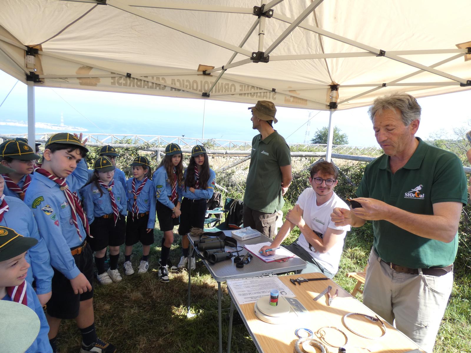
The full dataset is openly available on Zenodo, and the work is coordinated by the Stazione Ornitologica Calabrese (StOrCal), which brings the region’s observers together under shared standards. Much is still missing: the inland massifs of Pollino, Sila and Aspromonte remain under-surveyed, and breeding densities are poorly known for most species. The checklist gives that work a verifiable point to build on.

Original source:

Urso S, Martino G, Muscianese E, Policastrese M, Pucci M, Vena M, Storino P (2026) Updated checklist of the birds of Calabria (southern Italy). Biodiversity Data Journal 14: e195967. https://doi.org/10.3897/BDJ.14.e195967

A New Class Of Fungi Named After The King Of Sweden

A newly discovered fungal species has been named after the King of Sweden to mark the 50th anniversary of his reign.

Originally published by Uppsala University

Researchers at Uppsala University have discovered a completely new fungal species, which they chose to name after the King of Sweden. The species has been given the Latin name Semicentenialea rex, which means fifty-year anniversary of the King. It is the first known representative of a new class of fungi that will be called Semicentenialomycetes

The new fungus was isolated from roots in the ground in Jädraås in the province of Gästrikland, and represents a new lineage among what are primarily rust fungi. Rust fungi are a type of fungi that causes diseases in plants, usually by infecting the leaves. Based on DNA sequencing from soil samples, the researchers know that the class occurs in soil and roots from different ecosystems across almost the entire world, but rarely at high abundance.

the new fungi Semicentenialea rex
 The images show Semicentenialea rex both as a colony and under the microscope. After eight weeks of growth, the fungus forms a small colony on the culture plate. The microscopic images reveal a network of hyphae with so-called clamp connections – structures that connect adjacent cells in the hyphae of basidiomycete fungi. To follow fungal development, the researchers stained the nuclei pink and the cell walls blue. Three types of swollen cells can be observed within the hyphae: basidium-like cells, teliospore- or probasidium-like cells, and intercalary swellings. These structures are examined in greater detail in the following figures. Credit to Anna Rosling.

There are probably at least two other species in the class, but they do not appear to be as common as Semicentenialea rex. Since it only seems to live deep down in the ground, we are curious about what life strategy it has. We are going to study how it affects the roots of plants in the soil and how it propagates.

says Anna Rosling, professor at Uppsala University and one of the researchers behind the study.

Several million species of fungi

phylogenetic tree of Semicentenialea rex
The researchers analysed hundreds of genes to determine where the newly discovered fungus Semicentenialea rex belongs in the fungal tree of life. The results show that S. rex, previously known as GS25, forms a distinct lineage within the Pucciniomycotina. To confirm its placement, genes from both closely related and more distantly related fungi were compared, including species from both the Basidiomycota and the Ascomycota. Credit to Anna Rosling.

The species diversity in kingdom Fungi is largely unexplored. Based on sequencing of environmental DNA, it is estimated that there are several million species of fungi across the globe. But because most live concealed in the soil, wood or insects for example, it has only been possible to describe and name a small proportion of them. Without names, it is difficult to communicate about these species, genera, classes and even phyla of hitherto unknown fungi.

It was during a study root fungal diversity, when the researchers were culturing fungi from pine tree roots collected from mineral soil at Ivantjärnheden field station near Jädraås that the researchers identified a new fungal species that turned out to represent a new class of fungi.

Exciting with a root-associated fungus

Veera Tuovinen-Nogerius and Anna Rosling, both of Uppsala University, have led an international team of researchers in the work of characterising the morphology of the new species and its phylogenetic placement. The results have now been published in the International Mycology Association’s journal IMA Fungus.

A new class is exciting enough but in this case, it’s especially exciting because we have a root-associated fungus that evolutionarily speaking sits among the rust fungi in Puccinomycotina. Rust fungi are obligate parasites that infect plants and cause diseases above ground.

says Anna

Suitably named after the King of Sweden

This figure presents additional structures that are likely to play a role in the fungal life cycle. Among them are intercalary swellings and teliospore-like cells, which contain spore-like structures with one or more nuclei. The researchers documented several stages of development, ranging from young swellings and spore formation to germination and the emergence of new hyphae. Some structures produce thick promycelium-like hyphae, while others release small yeast-like cells through budding. These observations suggest that Semicentenialea rex has a complex life cycle involving several different cell types and spore forms. This resembles rust fungi within the Pucciniomycotina, which are known for producing multiple types of spores. Credit to Anna Rosling/Uppsala University.

Finding names for new species is a pleasant challenge for all researchers doing this type of research. This particular one was named Semicentenialea rex to pay tribute to King Carl XVI Gustaf of Sweden when he celebrated 50 years on the throne in 2023.

During his reign, the King has worked tirelessly for biodiversity conservation and the sustainable use of our natural resources. What could be better than to have a previously undescribed fungus named after you.

says Anna Rosling, who in connection with the official celebration of the King’s golden jubilee presented a picture and a description of the species to him.

Now that the species has a new name, it will facilitate future communication and studies of the role of Semicentenialea rex in the ecosystem.

Original source:

Nogerius VT, Sánchez-García M, Kluting K, Heinonsalo J, Ryberg M, Toome M, Haridas S, Mondo S, LaButti K, Nolan M, Lipzen A, Koriabine M, Bauer D, Barry K, Grigoriev IV, Aime MC, Rosling A (2026) Another dark taxon comes to light: Semicentenialomycetes, a new class within the Pucciniomycotina (Basidiomycota), and its first described representative, Semicentenialea rex. IMA Fungus 17: e189848. https://doi.org/10.3897/imafungus.17.189848

Camera Traps Reveal the True Culprit Behind Crop Damage in Honduras 

Using noninvasive monitoring in eastern Honduras, researchers found that small mammals, not large wildlife like tapirs, drive cassava crop loss, highlighting the need for evidence-based, wildlife-friendly solutions.

Guest blog post by Manfredo Turcios-Casco

Today, 29th June, marks the International Day of the Tropics, a day that reminds us of why understanding tropical ecosystems matters for both wildlife and the communities that depend on them. This new study from the Honduran Mosquitia is a perfect example. 

A new study from the Honduran Mosquitia shows how simple, non-invasive technology can help solve one of the most common challenges in wildlife conservation: identifying the species actually responsible for crop damage. The full detailed results have been published in the open-access peer-reviewed journal Neotropical Biology and Conservation.

Across tropical landscapes, people living near forests often share the same concern: wildlife entering agricultural fields and feeding on crops. In many cases, these interactions can generate tension between local communities and conservation efforts, particularly when threatened species are perceived as the main culprits.

Baird’s tapir (Tapirus bairdii) recorded from one of the trap cameras. Credit to the Wildlife Conservation Society (WCS).

In the Indigenous Miskitu community of Mavita, in eastern Honduras, local people have long reported losses in their cassava (Manihot esculenta) fields, locally known as yucales. Most residents believed that the damage was caused by Baird’s tapir (Tapirus bairdii), an endangered species and the largest terrestrial mammal in Central America. They also suspected that pacas (Cuniculus paca) and armadillos (Dasypus mexicanus) were contributing to crop losses.

But was the tapir really responsible?

To answer this question, researchers from the Wildlife Conservation Society (WCS) installed camera traps equipped with solar-powered motion-activated LED lights around a 10-hectare cassava field located within a mosaic of Caribbean pine forest and tropical rainforest in the Honduran Mosquitia.

Location of the motion-sensor light installed in the cassava field of Mavita, within the Mosquitia region of eastern Honduras, highlighting nearby communities (Mavita and Rus Rus) and the surrounding forest matrix based on the 2018 ICF land-cover classification. Credit to Turcios-Casco et al., 2026

The goal was to document which mammals were actually visiting the crops and evaluate whether these light systems could eventually help reduce crop losses. 

Following the evidence

Over two months of monitoring, the cameras recorded seven mammal species, including tapirs, ocelots, jaguarundis, agoutis, opossums and rabbits. 

Contrary to local perceptions, the species most frequently detected interacting with cassava crops was not the tapir, but the Honduran cottontail rabbit (Sylvilagus hondurensis) – a species that locals did not even know occurred in their plantations. The cameras also found no evidence that armadillos or pacas were feeding on the cassava.

Meanwhile, tapirs were present in the area, but appeared far less frequently than expected. 

Many conservation conflicts begin with assumptions. Without evidence, it is easy to blame large and conspicuous animals. Camera traps allowed us to identify which species were truly interacting with the crops and helped us separate perception from reality.

explains lead author Manfredo Turcios-Casco
@pensoft.publishers

On the #InternationalDayOfTheTropics , here’s a story that shows why understanding tropical ecosystems matters. 👇 📸What if the culprit wasn’t who we thought? Using noninvasive monitoring in eastern Honduras, researchers found that small mammals, not large wildlife like tapirs, drive cassava crop loss, highlighting the need for evidence-based, wildlife-friendly solutions. 📗Read the full study in Neotropical Biology and Conservation open-access journal: https://doi.org/10.3897/neotropical.21.e187958 👉Or check out our blog for more insights from the lead author and researcher, Manfredo Turcios-Casco: https://blog.pensoft.net/2026/06/25/camera-traps-reveal-the-true-culprit-behind-crop-damage-in-honduras/ @Wildlife Conservation Society #tapir #fyp

♬ A moist healing song – Nez Tunes

Why does this matter?

Correctly identifying the species involved in crop damage is more important than it might seem.

Across Latin America, wildlife is often persecuted after being blamed for agricultural losses. In Honduras, Baird’s tapir has historically faced retaliatory hunting in areas where farmers perceive it as a threat to their crops. 

When damage is incorrectly attributed to threatened species, conservation efforts can be undermined while the actual source of the problem goes unaddressed. 

The study highlights how non-invasive monitoring can provide communities and conservation practitioners with reliable information before management decisions are made. 

Technology for coexistence

The project also tested the use of solar-powered motion-sensor lights, sometimes referred to as “silent technology,” as a tool to monitor and potentially deter wildlife activity.

Researchers found that different species reacted differently to the lights. Tapirs tended to show stronger behavioral responses, while rabbits often continued moving through the area despite illumination. These findings suggest that no single deterrent works for every species and that mitigation strategies should be tailored to local ecological conditions. 

Solar-powered LED motion-sensor light interacting with female Tapirus bairdii (A), male Tapirus bairdii (B), and Sylvilagus hondurensis (C).
Solar-powered LED motion-sensor light interacting with female Tapirus bairdii (A), male Tapirus bairdii (B), and Sylvilagus hondurensis (C). Credit to Turcios-Casco et al., 2026

More importantly, the technology proved valuable as a diagnostic tool, helping researchers understand not only which species were present, but also when they were active and how they responded to human-made stimuli. 

Conservation begins with understanding

Solar-powered LED motion-sensor light interacting with Leopardus wiedii (above) and Didelphis marsupialis (below)
Solar-powered LED motion-sensor light interacting with Leopardus wiedii (above) and Didelphis marsupialis (below). Credit to Turcios-Casco et al., 2026

The forests of the Honduran Mosquitia harbor some of the most important wildlife populations remaining in Central America. Yet the long-term conservation of these species depends not only on protecting habitat, but also on fostering coexistence with local communities. 

What surprised me most was discovering that the species most frequently blamed by local people was not the one causing most of the crop interactions.

expressed Manfredo Turcios-Casco

This study demonstrates that effective conservation starts with understanding what is actually happening on the ground. Sometimes the evidence confirms what people already suspect. And sometimes it reveals that the animal everyone blamed was innocent all along. 

Acknowledgments
This article was produced by the Wildlife Conservation Society (WCS), with technical support of the community of Mavita and the financial support from the Fondo para el Manejo de Áreas Protegidas y Vida Silvestre (FAVPS) and the Biodiverse Landscapes Fund (BLF), funded by UK International Development. The views expressed do not necessarily reflect the official policies of the UK Government.

Original source

Turcios-Casco MA, Jolon-Morales MR, Padilla B, Scott E, López CM (2026) From forest mosaics to yucales: noninvasive monitoring untangles mammal–crop interactions in eastern Honduras. Neotropical Biology and Conservation 21(2): 173-188. https://doi.org/10.3897/neotropical.21.e187958

When Traded Frogs Are Not What They Seem: Hidden Identities Behind The Amphibian Trade

A new study published in Nature Conservation reveals that threatened amphibian species are being sold under incorrect names in online amphibian trade marketplaces.

Guest blog post by Xiaoli Zhang

When we first began this work, our original aim was to study the evolutionary history and divergence time of the genus Hoplobatrachus using genetic data. The traded specimens we collected were intended to support this phylogenetic analysis, not to investigate the legality of the amphibian trade. However, during samples verification, we found that some frogs sold as Quasipaa were genetically Hoplobatrachus, highlighting mismatch between trade labels and actual species identity.

This discovery led us to take a closer look at species identification in amphibian trade. Like many researchers in wildlife biology, we initially assumed that most trade labels were generally reliable, even if not perfect. What we found turned out to be more complicated and the results have been published in the open-access journal Nature Conservation.

A simple verification revealed unexpected results

Left: Quasipaa spinosa, right: Hoplobatrachus chinensis
Left: Quasipaa spinosa, right: Hoplobatrachus chinensis, photos by Amaël Borzée, via iNaturalist (https://www.inaturalist.org/), licensed under CC BY-NC.

Our study focused on frogs traded under the name Hoplobatrachus chinensis, a species commonly recorded in commercial markets. To verify their identity, we used genetic tools and the results were clear: specimens labelled as Chinese edible frogs (Hoplobatrachus chinensis) were in fact Chinese spiny frogs (Quasipaa spinosa), a different species entirely and one that is classified as Vulnerable by the IUCN. At first glance, this might appear to be a minor taxonomic issue. However, it raises an important question: how reliable is species identification in wildlife trade once animals enter real-world commercial systems?

Why do these mistakes happen?

One of the main challenges in amphibian trade is that many species are very similar in appearance. Even for experienced handlers, distinguishing between certain species based on morphology alone can be extremely difficult. In most trade contexts, identification is based solely on external characteristics, without genetic confirmation. This creates room for both unintentional misidentification and potential substitution errors. Once specimens move through multiple steps in complex supply chains, their original identity may become increasingly uncertain.

Is this an isolated case?

Based on our findings, we suspect this is not an isolated issue. Instead, our results likely represent only the “tip of the iceberg.” Amphibians are traded in large numbers, often through complex and poorly traceable supply chains. In such systems, even small identification errors can accumulate and go unnoticed. Because genetic verification is rarely applied in routine monitoring, similar cases may be more widespread than currently documented.

Why does it matter?

Quasipaa spinosa 
Four Quasipaa spinosa purchased online under the false visual of Hoplobatrachus chinensis, a farmed species. Panels correspond to individual specimens that were bought. Credit to Zhang et al., 2026

At first, species misidentification may seem like a technical detail. However, it has direct consequences for conservation and wildlife trade regulation. If traded individuals are incorrectly identified, trade data may not accurately reflect which species are being exploited. This can lead to biased assessments of population pressure and potentially mask risks for threatened species. In other words, what is recorded in trade databases may not always match biological reality.

What can be done?

One of the key implications of our study is the importance of incorporating molecular tools into wildlife trade monitoring. DNA-based identification provides a reliable way to confirm species identity, particularly in cases where morphology alone is insufficient. While it may not be feasible to genetically test every traded individual, targeted or random sampling could significantly improve the accuracy and transparency of trade data.

A broader perspective

Beyond amphibians, our findings highlight a more general issue in wildlife trade: hidden uncertainty in species identification can quietly distort our understanding of biodiversity exploitation. Improving traceability and verification is therefore not just a technical improvement, but an essential step toward more effective conservation decision-making.

Original source:

Zhang, X., Borzée, A., Gill, S., & Othman, S. N. (2026). Hidden or inadvertent illegal amphibian trade? Genetic analyses reveal Quasipaa spinosa misidentified as Hoplobatrachus chinensis in the trade. Nature Conservation, 63, 247-257. DOI: 10.3897/natureconservation.63.183849

Invasive Reeds Can Promote The Proliferation Of Mosquitoes In Rivers, Canals And Ponds

The accumulation of leaf litter from this invasive alien plants facilitates the development of the larvae of the common mosquito, a species of ecological and public health concern.

Guest blog post by Alberto Maceda-Veiga and Rosa Martínez

Reed, an invasive alien plant that is abundant on the banks of many rivers, ponds and canals, can encourage the growth of common mosquito populations in the absence of natural predators. When the plant’s litter accumulates, the chemical properties of the water and the composition of the biological communities in the environment change radically, and this facilitates the development of mosquito larvae in freshwater ecosystems.

This is one of the conclusions of a study published in the journal NeoBiota and led by Professor Alberto Maceda-Veiga, a member of the FORESTREAM Research Group at the Faculty of Biology and the Biodiversity Research Institute (IRBio) at the University of Barcelona.

The study, whose lead author is Oriol Cano-Rocabayera, involved collaboration from experts at the Institute for Agri-Food Research and Technology (IRTA-UAB), the Mosquito Control Service (SCM) of El Baix Llobregat Regional Council, the University of Seville, the Doñana Biological Station (EBD-CSIC), the Goethe University (Frankfurt, Germany), the University of Florida (United States) and the universities of Cardiff and Chester (United Kingdom).

More mosquitoes if the invasive reed is present

Schematic food web of the trophic cascade resulting from leaf litter decomposition and the interaction between microeukaryotes and mosquito larvae.
Schematic food web of the trophic cascade resulting from leaf litter decomposition and the interaction between microeukaryotes and mosquito larvae. Microeukaryotes can be prey, trophic competitors, and parasites and potentially alter the internal microbiota of mosquito larvae through changes in waterborne bacteria and fungi. Represented microeukaryotes are testate amoeba, a. Naked amoeba; b. Euglenid flagellate; c. Bodonid flagellate; d. Planktonic ciliate, e; crawling ciliates; f. Sessile ciliate; g. and micrometazoa (small metazoa): nematoda, h. and oligochaeta, i. Credit to Cano-Rocabayera et al., 2026.

The ecological impacts of invasive riparian plants at the ecosystem level are not yet fully understood, beyond their impact on specific groups of organisms or on environmental conditions. In addition to altering biodiversity and the composition of the natural environment, exotic and invasive riparian vegetation can affect human well-being.

In the study, the team used experimental aquariums (microcosms) to analyse the ecological impact of replacing native common reed (Phragmites australis) with giant reed (Arundo donax), a species classified as invasive and similar in appearance.

In the presence of A. donax, the mosquito larvae survived longer and grew larger and faster. Behind this increase lies the proportion of A. donax leaf litter, which altered the water’s physicochemical properties and the composition of populations of organisms known as microeukaryotes — protozoa, fungi and other microscopic organisms – which are essential to the functioning of the ecosystem.

The study highlights a dramatic ecological effect, even at low concentrations of A. donax compared with P. australis. “This effect was catalysed by changes in water quality and in the abundance of certain groups of microeukaryotes, such as flagellates and amoebae, which form part of the microbial food webs on which the larvae of the common mosquito Culex pipiens feed,” says Professor Alberto Maceda-Veiga, from the UB’s Department of Evolutionary Biology, Ecology and Environmental Sciences.

It is important to remember — he continues — that the common mosquito can act as a vector for diseases of medical and veterinary significance. Identifying which plants encourage the proliferation of mosquitoes helps us to understand the complex relationships that species form in nature and, in turn, helps pest control services to predict where they are most likely to find larvae and to apply control measures, where necessary, due to the risk to human health.

Improving environmental management along river courses

Giant reed
The giant reed, an exotic and invasive plant, is capable of altering the ecological balance in the natural environment. Photo credit to Alberto Maceda-Veiga.

The reed, which can withstand high summer temperatures, typically produces a large amount of biomass and is even being used to generate alternative energy sources. Interestingly, in previous studies, the team had described the potential positive effects of the species, as it provides shelter and protection for native fish in rivers with typically poor coverage of riparian vegetation (Science of The Total Environment, 2025).

Now, the findings of this study shed light on an unexpected aspect of the ecological impact of this invasive plant in riparian areas.

In freshwater ecosystems most severely affected by chemical pollution, the natural aquatic predators of the larvae – such as fish and dragonflies – are often absent. Many larvae mean many adult mosquitoes, which can cause nuisance to humans and even pose health risks.

In light of these new findings, the authors suggest prioritizing the initial eradication of A. donax in infested areas and restoring these sites with native vegetation such as P. australis, as well as encouraging future studies to determine the breeding preferences of mosquitoes in these habitats and their interactions with their natural predators.

When the eradication of an invasive species is not feasible, we turn to mitigation measures to reduce its impact. In the case of mosquitoes, we must not give in to alarmism. Firstly, we must remember that in nature, mosquitoes have many predators, as they are a natural part of food webs. Secondly, we have some fantastic mosquito control services, such as the Baix Llobregat SCM participating in this study, which ensure that mosquitoes do not become a problem in areas with high human traffic, such as cities and the marshes of peri-urban environments.

concludes Maceda-Veiga

Original source:

Cano-Rocabayera O, Bonilla YY, Salvadó H, Garcia-Bargalló M, Sobhy IS, Berry C, Vilà M, Sabater F, Aranda C, Maceda-Veiga A (2026) Invasive plant-induced shifts in water chemistry and microeukaryotes enhance mosquito development. NeoBiota 106: 265-286. https://doi.org/10.3897/neobiota.106.167149

Modelling Life Beneath Our Feet: A New Step Towards Realistic Soil Ecology At The Landscape Scale

New Formal Model in the open-access Agricultural and Environmental Modelling journal contributes to a more realistic soil ecology modelling at the landscape level.

Guest blog post by Liyan Xie

Why focus on soil organisms?

Agricultural landscape
Agricultural landscape. Image via Canva.

Soil health is a core priority of the EU Soil Strategy for 2030, and soil organisms like Collembola (springtails) play a fundamental role in sustaining it. With an estimated 100,000 individuals per square meter in healthy soils, these invertebrates drive essential processes like nutrient cycling, organic matter fragmentation, and microbial regulation, while also serving as crucial prey for predators like spiders and mites. Despite their ecological importance and their distinct chemical exposure pathways compared to earthworms, they remain underrepresented in landscape-level environmental risk assessments, which often rely on simplified laboratory studies.

Moving beyond simplified tests

Assessing multiple ecosystem stressors
A framework for assessing multiple ecosystem stressors. Image credit to Liyan Xie.

Traditional ecotoxicological testing, such as standard OECD laboratory tests, has provided valuable baseline data for over 60 years, but these tests are typically conducted under constant, highly controlled conditions. While informative, this approach overlooks the dynamic nature of environmental drivers like temperature and soil moisture, which jointly shape physiological performance.

For example, existing population models often ignore soil moisture entirely or rely on simplistic constant temperatures. Addressing this gap requires advanced modelling tools that can represent non-linear life-history processes, such as stage transitions and vital rates, under realistic, fluctuating conditions.

To achieve this, the researchers utilized the Animal, Landscape and Man Simulation System (ALMaSS), a spatially explicit modelling framework that integrates static landscape features like soil types with dynamic, daily components like hourly weather data, crop management practices, and vegetation growth to simulate realistic environments for species populations

A mechanistic model of a soil species

In a recently published Formal Model in the open-access Agricultural and Environmental Modelling journal, researchers developed a spatially explicit stage-structured population model for the springtail Folsomia candida within the ALMaSS framework. The model explicitly represents egg, juvenile, and adult life stages, linking development, reproduction, and survival to environmental conditions using empirically parameterized thermal performance curves and dose-response functions. 

A key highlight of the model is its advanced estimation of surface soil water potential, which integrates high-resolution ERA5 weather data, evapotranspiration, and physical soil properties. By grounding these processes in biological mechanisms and factoring in food quality, the model provides a highly realistic representation of how soil populations respond to their environment

Scaling up: from local processes to landscapes

Spatial and temporal populations modelling.
Spatial and temporal populations modelling. Image credit to Liyan Xie.

The model is spatially explicit, simulating populations across large agricultural landscapes, typically 10 km by 10 km, divided into detailed polygons and grid cells. 

Using daily time steps for simulations lasting up to five years, the model captures subpopulation dynamics at a high-resolution spatial scale of 100 square meters. It even incorporates realistic, density-dependent dispersal behaviors, triggering adult springtails to migrate to surrounding cells when population densities exceed 100,000 per square meter or when food availability is severely restricted

This allows local environmental conditions to shape subpopulations, which together determine broader population dynamics. Such an approach enables exploration of how small-scale processes propagate to landscape-level patterns, including population persistence and recovery.

A foundation for future risk assessment

Even though the current version of the model focuses purely on environmental stressors without chemical exposure, its flexible structure serves as a foundational framework that can be easily extended. By eventually integrating toxicity modules, the model will enable the investigation of population-level impacts driven by agrochemical usage, such as pesticides and fertilizers, across different European farming practices. This mechanistic framework improves the interpretation of standardised laboratory and higher-tier mesocosm tests, providing a crucial tool for assessing the impact of multiple stressors under environmentally realistic scenarios.

Why this matters

This work highlights the critical importance of developing robust, process-based ecological models before introducing the additional complexities of chemical stressors. By carefully balancing biological realism with empirical data availability, the model provides actionable outputs, such as population growth rates, spatial distributions, and population recovery times, which serve as essential indicators of relative environmental risk. Ultimately, models like this offer a pathway towards more scientifically grounded, realistic assessments of ecosystem health in a changing environment

Original source:

Xie L, Duan X, Norouzi S, de Jonge LW, Topping CJ (2026) Integrating spatial and environmental stressors in a population model of Folsomia candida (Collembola, Isotomidae): a Formal Model within ALMaSS framework. Agricultural and Environmental Modelling 8: e184962. https://doi.org/10.3897/aem.8.184962 

Dead Fish Shed More Intact Cells Into The Environment: A Promising Source For High-Resolution Genomic Analysis From Water

Study in Metabarcoding and Metagenomics investigated the availability of intact fish cells in water as a source of high-quality eDNA, using live and dead Rainbow Trout.

Guest blog post by Hiroki Yamanaka

Fish naturally release their DNA into surrounding waters, a phenomenon that has long powered environmental DNA (eDNA) analysis for detecting species without direct observation. Now, we are aiming for a leap beyond simple species identification toward fine-scale genomic analysis – a frontier that demands much higher-quality DNA samples.

Environmental DNA analysis as a biodiversity assessment tool

Environmental DNA (eDNA) analysis is a non-invasive technique that extracts and detects the genetic material of target species from environmental samples such as water and soil, and it has rapidly transformed biodiversity monitoring. Compared to traditional tools, like catch-and-release or visual surveys, this technique possesses remarkably higher sensitivity, demonstrating unparalleled utility in detecting rare, elusive, or invasive species. Because it is cost-effective and highly efficient, eDNA analysis has become a new biodiversity assessment tool for mapping species distribution on a broad scale.

However, the current primary objective of eDNA analysis largely remains at qualitative species identification – simply asking “what species are here?” – making it an urgent priority to advance the technique toward extracting detailed and much deeper ecological information.

The importance of the “individuality” of cells for further development

The next great frontier for eDNA analysis is its application to population genetics. To understand the health, evolutionary history, genetic diversity, inbreeding, or to identify hybrid individuals within specific populations, extracting high-quality genomic information across multiple genes (multilocus) is essential.

Chum salmon
Chum salmon. Photo credit to Jeff Duda, one of the co-authors of the study.

However, free-floating DNA in the environment (bulk eDNA) is rapidly degraded by microbial activity and environmental factors, leaving it highly fragmented. Furthermore, because it is a “genetic soup” mixed with shredded DNA from hundreds of different individuals, the loss of physical linkage – the “individuality” of the genetic material – remains the greatest roadblock.

To solve this, we focused on capturing “intact cells.” A physically undamaged cell membrane acts like a biological shield, protecting the fragile DNA inside from the harsh, degrading environment outside. By isolating these intact cells from environmental water and performing single-cell analysis, it would make it possible to read the complete, uncorrupted genetic fingerprint of a single individual. The study is now available in the open-access journal Metabarcoding & Metagenomics.

eDNA from dead fish is not just noise

To establish a method to specifically detect and quantify intact cells from environmental samples, we conducted a five-day laboratory experiment using live and dead Rainbow Trout (Oncorhynchus mykiss).

  • Experimental tanks used in the study. Photo by Hiroki Yamanaka, Ryukoku University.
  • Experimental tanks used in the study. Photo by Hiroki Yamanaka, Ryukoku University.

To distinguish the DNA inside intact cells from the free-floating, degraded DNA in the environment, we utilized propidium monoazide (PMA), a photoreactive DNA intercalating dye. PMA permeates into broken cell membrane and binds the DNA inside off the cell as well as the free-floating DNA outside of the cells; when exposed to strong visible light, it forms cross-links with DNA molecules that physically block subsequent PCR amplification. This effectively silences the “noise,” allowing us to selectively quantify only the pristine DNA protected inside intact cells with healthy membranes.

Rainbow Trout waiting for the introduction to the experimental tanks. Photo by Hiroki Yamanaka, Ryukoku University.

The results of this experiment were a revelation that flips conventional understanding in eDNA analysis. Historically, fish carcasses were viewed as a significant nuisance-biological noise that sheds massive amounts of DNA, causing false-positive detections or distorting biomass estimates.

But, this study demonstrated that carcasses release significantly higher concentrations of both total eDNA and PMA-resistant (intact-cell) eDNA compared to living fish. Particularly on Day 2 and Day 3 of the experiment, tanks containing carcasses yielded roughly 30 times more intact-cell DNA than tanks with live fish. In short, carcasses proved to be a promising source for the high-integrity cells that researchers desperately need.

Cell-by-cell analysis opens the door for the future

Counting and measuring Rainbow Trout used in the study. Photo by Hiroki Yamanaka, Ryukoku University.

This discovery presents a paradigm shift in our understanding of environmental sampling strategies for advanced genomic analysis. To acquire pristine genetic data, sampling waters during or immediately after known mass mortality events in nature-such as the natural die-offs after Pacific salmon migrate and spawn-could be a highly effective approach.

Access to perfectly preserved intact cells paves the way for a revolution in environmental monitoring. By integrating single-cell genomics with eDNA technology, we can overcome the loss of physical linkage that has plagued population genetics. Instead of looking at a mixed picture of DNA from multiple individuals, isolating individual cells for analysis makes it possible to compute exact inbreeding coefficients, map out local population structures, and identify hybrid species. This effectively bridges the gap between basic species detection and highly advanced ecological tracking, providing a technological foundation to deeply monitor the genetic health of entire ecosystems from just a few liters of water.

Limitations and further efforts required

Illustration of Double Helix DNA Structure
Illustration of Double Helix DNA Structure. Credit to Sumali Ibnu Chamid via Canva.

While this research provides pioneering insights, vital challenges requiring further validation remain. First, because this was a carefully controlled laboratory experiment using a single species (Rainbow Trout), further empirical studies involving a diverse array of taxa in complex, unpredictable natural environments are needed to ensure universal application.

Second, the technological tools – specifically the PMA dye method – require fine-tuning. Our observations confirmed that it sometimes struggles to fully suppress the PCR amplification of extracellular DNA, particularly when targeting the very short DNA fragments (short amplicons) often used in fish eDNA assays. Additionally, as a biological reality, DNA self-degradation by internal cellular enzymes continues even inside the cell. Thus, scientists still need to quantitatively evaluate and model the exact decay rates of DNA housed inside these protective cell membranes.

Despite these technical hurdles, the approach of harnessing the intact cells shed by organisms after they die brightly illuminates a highly promising path toward unlocking the full, high-resolution genetic story of our natural world with parallel development of single cell analysis on the cells from water shed by multicellular organisms.

Note: single cell analysis is commonly conducted on microbes which are unicellular organisms. However, single cell analysis on environment-medium derived multicellular organisms is very scarce for now. The main purpose of the current work was to determine a good timing / good location for cell collection to assure higher concentration of cells with high quality DNA to make the technical tests easier for the single cell analysis development on environmental samples.

Original source:

Yamanaka H, Hirohara T, Hoy MS, Chase DM, Duda JJ, Ostberg CO (2026) Live and dead fish shed different amounts of intact cells: Implications for advancing environmental DNA methodologies. Metabarcoding and Metagenomics 10: e177451. https://doi.org/10.3897/mbmg.10.177451