Sixteen new fungus species found hiding in Thailand’s flowers

Researchers have discovered sixteen new species of fungus living unnoticed inside garden and wild flowers across northern Thailand, more than quintupling the number of such fungi known to occur in the country.

Researchers have discovered sixteen new species of fungus living unnoticed inside garden and wild flowers across northern Thailand, more than quintupling the number of such fungi known to occur in the country.

The fungi belong to Aureobasidium, a genus of dark, yeast-like organisms found almost everywhere on Earth, from human skin to glacial ice. Several members of the group are already prized in industry for producing useful enzymes, fighting plant disease and even breaking down plastic. Yet despite their familiarity, little was known about the diversity within Thailand’s tropical flowers.

Researchers from Chiang Mai University and Kasetsart University collected 54 fungal strains from 31 species of flower during a 2024 survey, among them bougainvillea, butter daisy, and periwinkle. Using a combination of DNA analysis across five gene regions, microscopy and growth trials, the team distinguished sixteen entirely new species, alongside five already known species (four of which had never before been recorded in Thailand).

Professor Nakarin Suwannarach of Chiang Mai University.

“Our team was motivated to study Aureobasidium because it was one of the most frequently isolated fungal genera from our flower samples.

We observed considerable variation among the isolates in colony morphology and other phenotypic characteristics, which suggested that there might be much greater species diversity than previously recognised.”

Professor Suwannarach
Periwinkle. Credit: Nakarin Suwannarach.
Melampodium divaricatum. Credit: Nakarin Suwannarach.

Distinguishing the new species was far from straightforward. Many looked almost identical under the microscope, and the DNA markers usually relied upon proved unable to tell them apart.

“This highlighted the limitations of relying on morphology alone, as well as on commonly used molecular markers.

By integrating multiple loci with morphological, physiological and growth-temperature data, we were able to distinguish closely related species more accurately. This was particularly surprising because Aureobasidium is a well-known fungal genus, yet our results revealed substantial previously unrecognised diversity in Thailand.”

Professor Suwannarach
Aureobasidium melanogenum – melanized hyphae developing into chlamydospores. Credit: Nakarin Suwannarach.

Most of the new species take their names from the flowers in which they were found, including Aureobasidium bougainvilleae, A. catharanthi and A. plumeriae, while others honour the northern Thai locations where they were collected.

Two species, A. saisamorniae and A. savitreeae, were named in tribute to two of Thailand’s leading mycologists, Professor Saisamorn Lumyong and Professor Savitree Limtong.

“We wanted the names to tell a story, either about where these fungi came from or about the people who have helped advance mycological research in Thailand.”

Professor Suwannarach
Bougainvillea. Credit: Nakarin Suwannarach.

Beyond the taxonomy, the team says the findings matter for the future. A small number of Aureobasidium species have been linked to opportunistic infections, so having an accurate record of which species occur naturally will help scientists spot and respond to any change in their behaviour over time.

“This study demonstrates that multilocus phylogenetic analysis is not simply a tool for naming new species. It can reveal hidden fungal diversity and provide essential baseline information for understanding and monitoring fungal populations in the future.”

Professor Suwannarach

The study is published in the open access journal IMA Fungus.


Original source:

Senwanna C, Kodchasee P, Kathongthung C, Samarakoon MC, Khunnamwong P, Kumla J, Suwannarach N (2026) Unexpected diversity of flower-associated Aureobasidium reveals sixteen novel species from northern Thailand. IMA Fungus 17: e188009. https://doi.org/10.3897/imafungus.17.188009

Pensoft Joins the Global Lichenology Community at IAL10 in Trieste

The 10th Symposium of the International Association for Lichenology took place in Trieste, where Pensoft showcased its plant science and mycology journal portfolio.

Last week, the 10th Symposium of the International Association for Lichenology (IAL10) took place in Trieste, Italy, from 26 to 31 July 2026, organised by the International Association for Lichenology, the University of Trieste’s Department of Life Sciences and the Italian Lichenological Society.

Held once every four years, the symposium is an important event for the global lichenology community, and this edition proved more popular than ever, drawing over 350 participants from around the world – more than double the number originally expected.

Lichenology is a wonderfully niche field, but IAL10 showed just how far-reaching and collaborative the community is. Delegates travelled from every corner of the globe to discuss taxonomy, lichen-associated microorganisms, biodiversity and conservation, ecophysiology, environmental biomonitoring, and cultural heritage preservation. 

Pensoft’s stand at IAL10

We were pleased to have our own stand at the symposium, where we showcased our journals in plant science and mycology, including:

  • MycoKeys: publishes papers on the monophyletic kingdom Fungi containing taxonomic or ecological data on any taxon of any geological age from any part of the world with no limit to manuscript size;
  • IMA Fungus: considers contributions from all areas of mycology expected to be of interest to the wider mycological community, from basic research to applications
  • PhytoKeys: publishes papers in systematic Botany containing taxonomic/floristic data on any taxon of any geological age from any part of the world 
  • Italian Botanist: publishes original research covering all fields of Botany in its wider sense, including Mycology, ranging from molecular to ecosystem studies
  • Biodiversity Data Journal: publishes papers in biodiversity science containing taxonomic, floristic/faunistic, morphological, genomic, phylogenetic, ecological or environmental data on any taxon of any geological age from any part of the world 
  • Vegetation Classification and Survey: publishes original papers that develop new vegetation typologies as well as applied studies that use such typologies, for example, in vegetation mapping, ecosystem modelling, nature conservation, land use management or monitoring 

It was a great opportunity to speak directly with researchers and students about publishing their work, and to reconnect with authors and editors already part of our community.

Talks that caught our attention

Among the many excellent presentations at IAL10, two in particular stood out to us:

Juri Nascimbene, a subject editor and author at MycoKeys and an author for Italian Botanist, Vegetation Classification and Survey, and Biodiversity Data Journal, presented on the historical lichen collections held at the University of Bologna’s herbarium (BOLO). His talk explored how these collections, some dating back to the work of Ulisse Aldrovandi in the sixteenth century, allow researchers to track species distributions across centuries. 

Using material such as Antonio Bertoloni’s Hortus Siccus Florae Italicae, which contains 850 georeferenced lichen specimens, Nascimbene’s team has been able to trace the long-term presence of species such as Lobaria pulmonaria in the chestnut groves of the Northern Apennines, and the disappearance of cold-adapted species such as Alectoria ochroleuca from high-altitude sites.

It was a fascinating look at how historical herbaria can inform our understanding of global change today!

We also attended a talk by Martin Grube, a reviewer and author at MycoKeys, who challenged the common perception of lichens as slow-growing organisms. His presentation examined the hygroscopic movements lichens undergo as they take up and lose water, and the mechanical stresses this creates within their thalli.

Grube introduced tools for measuring these mechanical properties and discussed what they might reveal about the evolution and function of lichen structures, thus offering a compelling primer on comparative thallus mechanics.

A new special issue in MycoKeys

While at IAL10, we were pleased to announce a new special issue in MycoKeys: Proceedings of the 10th International Conference of the International Association for Lichenology (IAL10), edited by Mauro Tretiach, Fabio Candotto Carniel, and Lucia Muggia. The special issue will bring together original research articles and short communications covering the full breadth of lichen symbiosis research. We look forward to seeing the community’s latest work published there.

Celebrating the award winners

Congratulations to all of this year’s IAL10 award winners:

Group photograph of the award winners at IAL10.

Best Posters

  • Honorata Wacławek, “Phylogenetic insights into freshwater Gyalidea (Gomphillaceae, Graphidales) from the high Andes of Bolivia”
  • Ariel Sebastián Udaeta Sanchez, “First axenic cultures of saxicolous lichens from Tucumán (NW Argentina): growth timelines and representation of under-studied neotropical lineages”

Best Flash Talks

  • Ciaran Kelly, “Comparative genomic analyses of antimicrobial proteins encoded by lichen-forming fungi”
  • Anna Pasinato, “The metabolic divide: fungal genomes reveal an unequal distribution of biosynthetic gene clusters”

Best Oral Presentations

  • Victoria K., “Hidden cohabitants: metagenomic insights into global occurrence patterns of black fungi associated with lichens”
  • Diego Garfias Gallegos, “Central metabolism and development are rewired in lichenized cyanobacteria”

Until next time

IAL10 was a wonderful opportunity to reconnect with the lichenology community, and we were delighted to meet so many authors from our journals in person. Thank you to everyone who stopped by our stand, attended a talk with us, or simply shared a conversation about lichens.

We look forward to continuing our collaboration with the Italian Lichenological Society and seeing you all again at the next symposium. 


Follow us on Bluesky, Facebook, X, and LinkedIn for future conference updates.

Amazonian ‘zombie’ fungus harbours a hidden stage inside forest mosses, new study reveals

Research led by INPA scientists reveals that the parasite, famous for altering ant behavior, also shelters within vegetation, suggesting a dual ecological journey never before observed in manipulating lineages of the genus.

A team led by researchers at the National Institute for Amazonian Research (INPA), in Manaus, has found evidence that the “zombie-ant” fungus (genus Ophiocordyceps), globally known for controlling ant behavior and forcing infected ants to bite onto plants before dying, also lives hidden inside mosses in the Amazon rainforest.

By analysing the DNA of mosses collected at the Ducke Reserve in Manaus, the scientists detected the same parasitic fungus present not only in infected and manipulated insects, but also in the mosses where the infected insect is manipulated to bite. This indicates that the fungus has one life stage as an insect parasite and another, endophytic (living inside the plant) stage that had gone unnoticed until now.

The genus Ophiocordyceps comprises more than 350 species of fungi known for infecting a wide range of insects. Some of these species are famous for a behaviour that sounds like science fiction: after infecting an ant, the fungus alters its behaviour and forces it to leave the colony, climb the vegetation, and clamp its mandibles onto a specific spot, the so-called “death grip,” shortly before dying and releasing its spores.

In 2020, studies in China had already shown that a species from the same genus, known as “Himalayan gold” (O. sinensis), lived endophytically in plants in alpine regions. The major breakthrough of this new INPA study is demonstrating this same dual lifestyle in fungi that manipulate the insect to specifically and consistently bite plants that harbor the endophytic stage of the “zombie fungus.”

This suggests that the behavioral manipulation goes beyond the fungus-ant association, and that the plant harbouring the fungus’ endophytic phase may also possibly influence the manipulation itself.

The research, published in the open-access journal IMA Fungus, was carried out at the Adolpho Ducke Forest Reserve, north of Manaus, and relied on metabarcoding, a technique that allows researchers to read the DNA of all fungi present in an environmental sample at once.

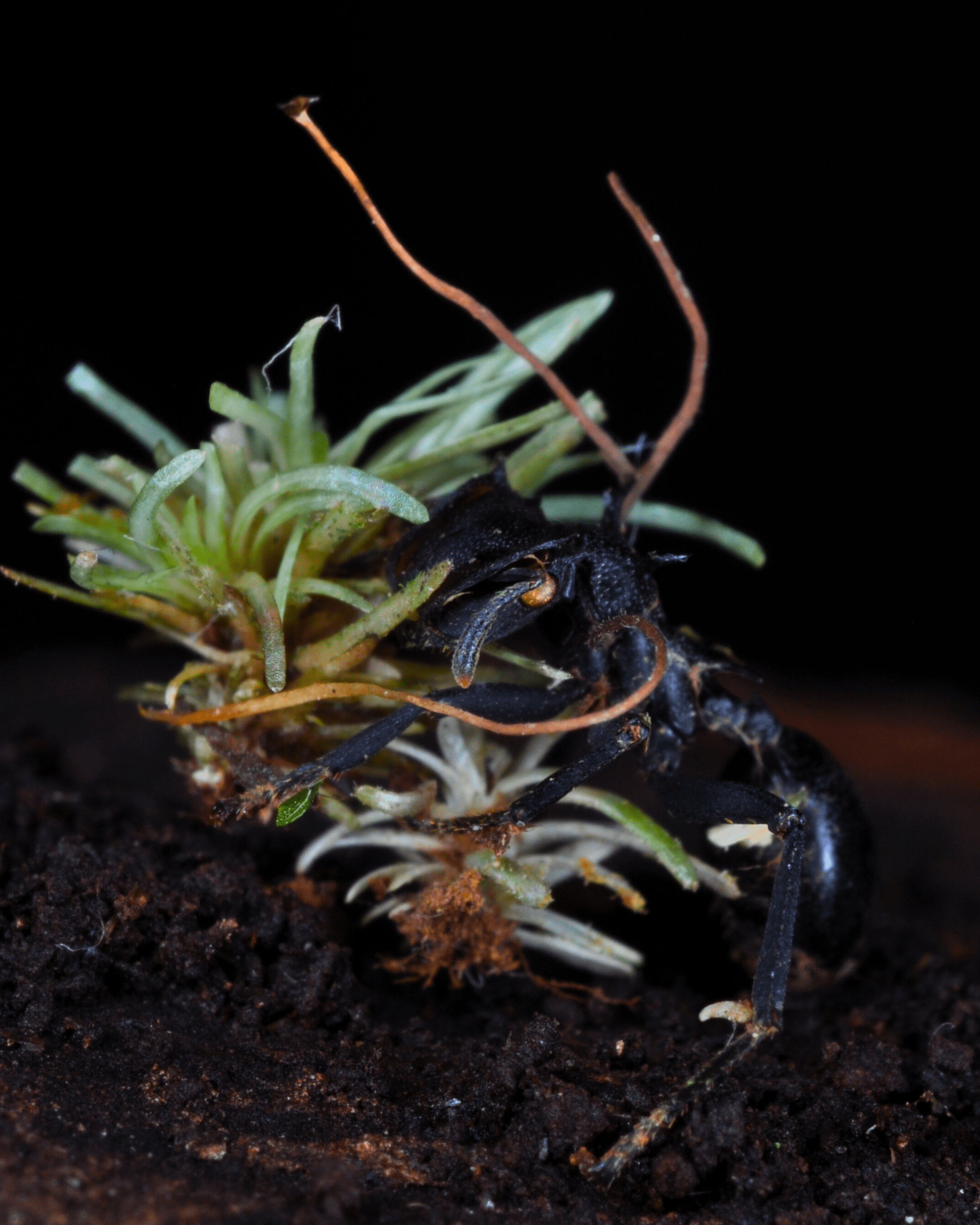
Ophiocordyceps-infected insects found attached to bryophytes. Camponotus renggeri ant infected by O. camponoti-renggeri (TAJ32) biting into a Plagiochila liverwort while gripping an Octoblepharum moss with its tarsal claws. Image credit: Tales Alves-Júnior et al.

The results revealed that the fungus was present not only in infected ants but also consistently in the surrounding mosses, including in plants located more than 10 metres away from any ant infected by the parasite.

In the Amazon, researchers had already noticed that some manipulated ant species predominantly seek out mosses to bite for the “death grip,” but little was known about a possible prior colonisation of the moss by the same insect-parasitising fungus.

DNA analyses showed that even highly host-specialised fungal lineages, such as O. camponoti-nidulantis (infecting Camponotus nidulans) and O. kniphofioides (infecting Cephalotes atratus), were sheltered within bryophytes in the surrounding vegetation.

Ophiocordyceps kniphofioides in a turtle ant (Cephalotes sp.), found attached to an Octoblepharum moss. Image credit: Tales Alves-Júnior.

The discovery reshapes our understanding of the life cycle of these fungi and how these organisms evolve and survive. The ability to associate with plants may help the fungus persist through periods when insect hosts are scarce, and could also help explain the intriguing fidelity of certain fungal lineages to specific moss types.

“We have always thought of this fungus (Ophiocordyceps) as a parasite exclusive to insects, especially the ones it manipulates. What our data suggest is that the story is much larger: the same fungus appears to be ubiquitous in the surrounding moss communities. Our results illustrate a far closer link between the lifecycles of the plants and fungi involved in the behavioral manipulation of Amazonian ants.

Tales Alves Jr., biologist, Master’s in Botany from INPA and lead author of the study.

“The well-known behavioral manipulation caused by Ophiocordyceps may not result solely from the interaction between fungus and ant, but from a complex relationship among the fungus, its host, and the plant where the same fungus lives silently as an endophyte.”

João Paulo Machado de Araújo, co-author and researcher at the Natural History Museum of Denmark (University of Copenhagen).

Original source

Alves-Júnior T, Araújo JPM, Góes-Neto A, Mendes-Pereira T, Zartman CE (2026) Metabarcoding reveals a hidden endophytic stage of the ‘zombie-ant’ fungus in Amazonian mosses. IMA Fungus 17: e196998. https://doi.org/10.3897/imafungus.17.196998

A New Class Of Fungi Named After The King Of Sweden

A newly discovered fungal species has been named after the King of Sweden to mark the 50th anniversary of his reign.

Originally published by Uppsala University

Researchers at Uppsala University have discovered a completely new fungal species, which they chose to name after the King of Sweden. The species has been given the Latin name Semicentenialea rex, which means fifty-year anniversary of the King. It is the first known representative of a new class of fungi that will be called Semicentenialomycetes

The new fungus was isolated from roots in the ground in Jädraås in the province of Gästrikland, and represents a new lineage among what are primarily rust fungi. Rust fungi are a type of fungi that causes diseases in plants, usually by infecting the leaves. Based on DNA sequencing from soil samples, the researchers know that the class occurs in soil and roots from different ecosystems across almost the entire world, but rarely at high abundance.

the new fungi Semicentenialea rex
 The images show Semicentenialea rex both as a colony and under the microscope. After eight weeks of growth, the fungus forms a small colony on the culture plate. The microscopic images reveal a network of hyphae with so-called clamp connections – structures that connect adjacent cells in the hyphae of basidiomycete fungi. To follow fungal development, the researchers stained the nuclei pink and the cell walls blue. Three types of swollen cells can be observed within the hyphae: basidium-like cells, teliospore- or probasidium-like cells, and intercalary swellings. These structures are examined in greater detail in the following figures. Credit to Anna Rosling.

There are probably at least two other species in the class, but they do not appear to be as common as Semicentenialea rex. Since it only seems to live deep down in the ground, we are curious about what life strategy it has. We are going to study how it affects the roots of plants in the soil and how it propagates.

says Anna Rosling, professor at Uppsala University and one of the researchers behind the study.

Several million species of fungi

phylogenetic tree of Semicentenialea rex
The researchers analysed hundreds of genes to determine where the newly discovered fungus Semicentenialea rex belongs in the fungal tree of life. The results show that S. rex, previously known as GS25, forms a distinct lineage within the Pucciniomycotina. To confirm its placement, genes from both closely related and more distantly related fungi were compared, including species from both the Basidiomycota and the Ascomycota. Credit to Anna Rosling.

The species diversity in kingdom Fungi is largely unexplored. Based on sequencing of environmental DNA, it is estimated that there are several million species of fungi across the globe. But because most live concealed in the soil, wood or insects for example, it has only been possible to describe and name a small proportion of them. Without names, it is difficult to communicate about these species, genera, classes and even phyla of hitherto unknown fungi.

It was during a study root fungal diversity, when the researchers were culturing fungi from pine tree roots collected from mineral soil at Ivantjärnheden field station near Jädraås that the researchers identified a new fungal species that turned out to represent a new class of fungi.

Exciting with a root-associated fungus

Veera Tuovinen-Nogerius and Anna Rosling, both of Uppsala University, have led an international team of researchers in the work of characterising the morphology of the new species and its phylogenetic placement. The results have now been published in the International Mycology Association’s journal IMA Fungus.

A new class is exciting enough but in this case, it’s especially exciting because we have a root-associated fungus that evolutionarily speaking sits among the rust fungi in Puccinomycotina. Rust fungi are obligate parasites that infect plants and cause diseases above ground.

says Anna

Suitably named after the King of Sweden

This figure presents additional structures that are likely to play a role in the fungal life cycle. Among them are intercalary swellings and teliospore-like cells, which contain spore-like structures with one or more nuclei. The researchers documented several stages of development, ranging from young swellings and spore formation to germination and the emergence of new hyphae. Some structures produce thick promycelium-like hyphae, while others release small yeast-like cells through budding. These observations suggest that Semicentenialea rex has a complex life cycle involving several different cell types and spore forms. This resembles rust fungi within the Pucciniomycotina, which are known for producing multiple types of spores. Credit to Anna Rosling/Uppsala University.

Finding names for new species is a pleasant challenge for all researchers doing this type of research. This particular one was named Semicentenialea rex to pay tribute to King Carl XVI Gustaf of Sweden when he celebrated 50 years on the throne in 2023.

During his reign, the King has worked tirelessly for biodiversity conservation and the sustainable use of our natural resources. What could be better than to have a previously undescribed fungus named after you.

says Anna Rosling, who in connection with the official celebration of the King’s golden jubilee presented a picture and a description of the species to him.

Now that the species has a new name, it will facilitate future communication and studies of the role of Semicentenialea rex in the ecosystem.

Original source:

Nogerius VT, Sánchez-García M, Kluting K, Heinonsalo J, Ryberg M, Toome M, Haridas S, Mondo S, LaButti K, Nolan M, Lipzen A, Koriabine M, Bauer D, Barry K, Grigoriev IV, Aime MC, Rosling A (2026) Another dark taxon comes to light: Semicentenialomycetes, a new class within the Pucciniomycotina (Basidiomycota), and its first described representative, Semicentenialea rex. IMA Fungus 17: e189848. https://doi.org/10.3897/imafungus.17.189848

From Spider Silk to Science: A New Way to Access Hidden Fungal Diversity

A new study suggests that spider webs can serve as natural, non-destructive collectors of fungal material in agricultural ecosystems.

A new study published in the open-access Biodiversity Data Journal suggests that spider webs – particularly those incorporating environmental debris – can serve as natural, non-destructive collectors of fungal material in agricultural ecosystems. The findings show that viable fungi can be recovered from these structures, including lineages that may represent previously undocumented diversity.

“Spider webs are often overlooked structures in the environment, yet they can function as natural collectors of biological material. Our findings suggest that they can be used as a complementary approach to access microbial communities without disturbing the surrounding ecosystem.”

Thanakron Into (Student at Thammasat University)

Researchers from Thammasat University and the National Center for Genetic Engineering and Biotechnology (BIOTEC), Thailand, investigated whether the adhesive and particle-trapping properties of spider silk could be used to capture and culture fungi associated with airborne and environmental particles. Unlike DNA-based methods, which detect genetic material regardless of viability, this approach allows for the recovery of living organisms that can be further studied.

The study focused on tropical rice fields, using webs of the orb-weaving spider Cyclosa mulmeinensis, a species known for constructing distinctive “trashline” decorations – linear accumulations of plant fragments, insect remains, and other debris within the web. These structures can intercept a variety of particles, some of which may carry fungal propagules.

Representative orb webs of Cyclosa mulmeinensis illustrating web architecture and debris decoration. Image credit: Thanakron Into et al.

Webs were collected from rice-field embankments in Pathum Thani, Nakhon Nayok, and Phetchaburi provinces using sterile techniques. In the laboratory, material retained on the silk was gently removed and cultured, yielding 112 viable fungal isolates. These isolates were grouped into 23 taxa across six genera, including Alternaria, Aspergillus, Cladosporium, Fusarium, Penicillium, and Talaromyces.

“We were particularly surprised that many of the fungi recovered from the webs remained viable and could be cultured. This enables further investigation beyond presence or absence, including their biological characteristics.”

Thanakron Into
Locations of paddy fields where Cyclosa mulmeinensis spider webs were collected in Thailand (left). Composition of culturable fungal taxa across genera in individual sampled webs, expressed as the number of taxa recovered per web (right). Image credit: Thanakron Into et al.

Some genetic lineages – particularly within Cladosporium and Talaromyces – did not match currently described species in available databases, indicating that additional, undocumented diversity may be present in these systems.

Conventional approaches to fungal monitoring typically rely on soil, air, or plant sampling, or on molecular methods that may not distinguish between living and non-living material. In this context, spider webs may provide a useful supplementary sampling surface for capturing biologically relevant particles.

Because spider webs are naturally maintained and, in some species, periodically rebuilt, this method can be applied with minimal disturbance to both the organisms and their environment. Importantly, the spiders themselves were not harmed during sampling, as only small sections of the web were collected.

“The ability to recover living fungi from these naturally occurring structures adds a practical dimension to biodiversity studies. It provides a way to link environmental sampling with downstream biological work.”

Thanakron Into

The idea that something as familiar as a spider web could quietly capture a hidden layer of biodiversity highlights how much of the natural world remains overlooked in plain sight.

While further work is needed to evaluate how broadly this approach can be applied, the study demonstrates the potential of spider webs as an additional tool for exploring microbial diversity in agricultural landscapes. 

Diagram displaying an overview of the study. Image credit: Booppa Petcharad.

Original source:

Into T, Petcharad B, Boonyuen N, Chanklan R, Pannanusorn S, Mongkolsamrit S, Kobmoo N, Nuankaew S, Kwanthong P (2026) Spider webs as reservoirs of culturable fungal diversity: evidence from orb-weaving Cyclosa mulmeinensis spider in Thai rice agroecosystems. Biodiversity Data Journal 14: e187035. https://doi.org/10.3897/BDJ.14.e187035

For more interesting articles on biodiversity, follow Biodiversity Data Journal on Facebook, Bluesky and X.

Inside the Hidden World of Spider-Attacking Fungi

Newly discovered groups of “zombie” fungi have been found to mummify spiders and adapt their physical forms.

Deep within the humid leaf litter of China and the dense canopies of Brazil’s Atlantic Forest, a silent ambush unfolds. 

While we often think of spiders as the ultimate predators of the undergrowth, they have an arch-nemesis: araneopathogenic fungi. These “zombie” fungi are capable of parasitising spiders by hijacking their bodies and consuming them from the inside out.

Two studies published in the open-access peer-reviewed scientific journals MycoKeys and IMA Fungus, respectively, offer insight into this macabre world of spider assassins.

In Southeast Asia, researchers led by Chen-xin Chang of the Guizhou University of Traditional Chinese Medicine have identified three new species of Gibellula fungi in China and Laos, which erupt from spiders in branch-like structures: Gibellula pseudopigmentosa, Gibellula pseudosolita, and Gibellula sinensis. These species are distinguished from one another by their slight variations in sexual reproductive structures and morphology. The below figure displays morphological plates, showing the three fungi species at both a macroscopic and microscopic level – notice their unique conidial heads and spore arrangements, coloured in blue.

To identify new species of spider-pathogenic fungi, the research group conducted field surveys in the forest leaf of China and Laos, where they collected specimens for detailed laboratory study. A combination of traditional microscopy and modern DNA sequencing rendered the discovery possible. The fungi’s sighting in Laos is particularly significant because it provides the first formal record of the Gibellula genus in the region. 

This study – published in MycoKeys – therefore serves to fill a major distributional gap in Southeast Asia, as well as expand our understanding of the morphological diversity within this group of spider-pathogenic fungi.

Meanwhile, in Brazil, a study led by Joao Paulo Machado De Araújo of the University of Copenhagen and the Royal Botanic Gardens of Kew, published in IMA Fungus, described a new species of Purpureocillium fungus belonging to the Purpureocillium atypicola group: Purpureocillium atlanticum. This fungus specifically targets trapdoor spiders inhabiting burrows on the forest floor, where it mummifies the host in white mycelia and subsequently emerges from its cephalothorax in the form of a purple fruiting body. 

The discovery was notably featured in The Guardian, where it was placed alongside other unusual botanical and fungal discoveries compiled by the Royal Botanic Gardens, Kew.

Phylogeny of hypocrealean fungi (A), highlighting the Purpureocillium atypicola complex (B) and morphology of the new species, P. atlanticum (C–G). Photo credit: Araújo et al.

Purpureocillium atypicola was originally recorded in Japan by Yasuda (1894), and was thought to be a single species found all over the world for over a century. The discovery of the Purpureocillium atlanticum in Brazil is significant because it finally confirms that Purpureocillium atypicola is actually a global complex of many unique species. 

To identify this new fungus, De Araújo’s research group used taxogenomics, a method which entailed bringing portable DNA sequencing gear directly into the Brazilian rainforest. By analysing the genetic code of the fungus and its environment immediately in the field, they were able to identify the specimen within just four days as opposed to waiting months for traditional lab results.

Both of these studies highlight the impressive diversity of spider-pathogenic fungi across distinct global environments. They additionally reveal the different evolutionary strategies of their respective species – while Purpureocillium atlanticum has adapted to infect underground trapdoor spiders by producing purple stalks to escape burrows, the Gibellula species represent the most diverse genus of spider parasites, found primarily in forest debris. 

As researchers continue to map these complex ecological networks, it becomes clear that preserving threatened biomes, including the Atlantic Forest and the jungles of Southeast Asia, is critical to expanding our knowledge in fungal taxonomy.

Original studies: 

Araújo JPM, Przelomska NAS, Smith RJ, Drechsler-Santos ER, Alves-Silva G, Martins-Cunha K, Hosoya T, Luangsa-ard JJ, Perrigo A, Repullés M, Matos-Maraví P, Woods R, Pérez-Escobar OA, Antonelli A (2025) A new species of Purpureocillium (Ophiocordycipitaceae) fungus parasitizing trapdoor spiders in Brazil’s Atlantic Forest and its associated microbiome revealed through in situ “taxogenomics”. IMA Fungus 16: e168534. https://doi.org/10.3897/imafungus.16.168534

Chang C-xin, Chen H, Loinheuang C, Dai Y-dong, Wang Y (2026) Morphological and phylogenetic analyses reveal three new species of Gibellula (Cordycipitaceae, Hypocreales) from spiders. MycoKeys 127: 135-154. https://doi.org/10.3897/mycokeys.127.177871

To stay up-to-date with new studies made openly available from the MycoKeys and IMA Fungus journal, subscribe to their journal newsletters via Email Alert on their journal websites, and don’t forget to follow them on social media!

Using AI to Uncover the Secret Lives of Fungi

AI holds the potential to automatically identify fungal versatility from the available scientific literature.

Fungi are the hidden architects of our ecosystems, acting as everything from helpful partners for plants to aggressive decomposers that recycle dead wood. However, many fungi don’t stick to just one job; they can switch lifestyles depending on their environment.  

Understanding this flexibility is vital for predicting how forests and farms will react to climate change. Unfortunately, the information researchers need is buried in decades of scientific papers that would take too long to comb through manually. 

A new study led by Northern Arizona University doctoral student Beatrice M. Bock and published in the open-access journal Research Ideas and Outcomes demonstrates how AI can solve this problem. By using a specialized language model called BioBERT, Bock developed an automated workflow that assesses scientific abstracts and accurately identifies whether a fungus has a single lifestyle or a dual, flexible one. 

A high-accuracy hack 

Bock said that for years, mycologists have relied on manual databases to track what different fungi do in the environment. While these tools are essential, they are difficult to keep updated as new research is published every day. 

“Manually identifying fungal versatility from the literature is time-consuming and difficult to scale. By using machine learning, we can now scan thousands of papers in just a few minutes to flag species that might be switching roles—such as a fungus that normally helps a plant grow but also turns into a decomposer when the plant dies.” 

Beatrice M. Bock

The pilot study tested four different AI models to see which was best at understanding the nuances of biological language. The top-performing model, BioBERT, achieved nearly 90% accuracy in identifying fungal lifestyles. 

What did BioBERT have that the other models didn’t? For one, it had the power of capitalization. Bock found that “cased” models—those that recognize capital letters—performed significantly better than those that did not. That’s likely because capital letters often signal species’ scientific names, like Fusarium, which are crucial for AI to understand the context of the research. 

The path ahead 

Bock said that in a commitment to transparency, she has made all the code and data available for free online, allowing other scientists to build upon her work and track traits in other organisms, like insects or plants. 

While Bock’s study focused on a small group of papers as a proof-of-concept, it opens the door for much larger projects. Future versions of the tool could predict how a fungus’s behavior might change under specific environmental conditions, such as drought or extreme heat. 

“As fungal trait databases continue to grow in importance for biodiversity assessments, automated text mining offers a path toward more efficient, consistent and comprehensive trait annotation.”

Beatrice M. Bock

Original sources:

Bock B (2026) Automated extraction of fungal trophic modes from literature using BioBERT: an open pilot workflow. Research Ideas and Outcomes 12: e176590. https://doi.org/10.3897/rio.12.e176590

Story originally published by: EurekAlert! (2026). Using AI to uncover the secret lives of fungi. [online] Available at: https://www.eurekalert.org/news-releases/1114462 [Accessed 2 Feb. 2026]. Republished with permission.

Follow the latest publications and news from the Research Ideas and Outcomes (RIO) journal on Bluesky, Facebook and LinkedIn.

Life on Mars? Lichens survive Martian simulation

Previously presumed uninhabitable, the Martian surface may be suitable for certain lichen species.

A thin atmosphere, freezing temperatures, and a barrage of radiation: the surface of Mars is hardly a prime holiday destination. But can any life survive there?

Known for their extreme tolerance to harsh environments such as Earth’s deserts and polar regions, lichens have long been considered a leading candidate for Martian survival. And, for the first time, researchers have demonstrated that certain species can survive Mars-like conditions, including exposure to ionising radiation, while maintaining a metabolically active state.

Published in the open-access journal IMA Fungus, a new study highlights the potential for lichens to survive and function on the Martian surface, challenging previous assumptions that the planet is uninhabitable.

Annotated design showing the set up of the experiment subjecting lichens to Mars-like conditions.
Experiment arrangement of vacuum chamber with the additional facility, including metal grate with lichens, cooling table, temperature, pressure and humidity sensors, X-ray lamp with the controller, CO2 valve with cylinder, controllers of vacuum chamber, pressure, cooling table, and computer.

But what exactly are lichens? It’s a little complicated. In fact, lichens are not a single organism, but rather a symbiotic association between a fungus and algae and/or cyanobacteria.

In this study, the fungal partner in lichen symbiosis remained metabolically active when exposed to Mars-like atmospheric conditions in darkness, including X-ray radiation levels expected on Mars over one year of strong solar activity.

Macro photograph of a lichen species.
Cetraria aculeata.

The research focuses on two lichen species (yes, there are lichen species despite them being a symbiosis), Diploschistes muscorum and Cetraria aculeata, selected for their differing traits. The lichens were exposed to Mars-like conditions for five hours in a simulation of the planet’s atmospheric composition, pressure, temperature fluctuations, and X-ray radiation.

The findings suggest that lichens, particularly D. muscorum, could potentially survive on Mars despite the high doses of X-ray radiation associated with solar flares and energetic particles reaching the planet’s surface. These results challenge the assumption that ionising radiation is an insurmountable barrier to life on Mars and set the stage for further research on the potential for extraterrestrial microbial and symbiotic survival.

“Our study is the first to demonstrate that the metabolism of the fungal partner in lichen symbiosis remained active while being in an environment resembling the surface of Mars. We found that Diploschistes muscorum was able to carry out metabolic processes and activate defense mechanisms effectively. 

“These findings expand our understanding of biological processes under simulated Martian conditions and reveal how hydrated organisms respond to ionizing radiation – one of the most critical challenges for survival and habitability on Mars. Ultimately, this research deepens our knowledge of lichen adaptation and their potential for colonizing extraterrestrial environments.”

Lead author of the paper, Kaja Skubała.

Further long-term studies investigating the impact of chronic radiation exposure on lichens have been recommended, as well as experiments assessing their survival in real Martian environments. 

Researchers from Jagiellonian University and the Space Research Centre of the Polish Academy of Sciences conducted the study with support from the National Science Centre, Poland, and the “Excellence Initiative – Research University” at the Faculty of Biology, Jagiellonian University.

Original study

Skubała K, Chowaniec K, Kowaliński M, Mrozek T, Bąkała J, Latkowska E, Myśliwa-Kurdziel B (2025) Ionizing radiation resilience: how metabolically active lichens endure exposure to the simulated Mars atmosphere. IMA Fungus 16: e145477. https://doi.org/10.3897/imafungus.16.145477

Follow IMA Fungus on Bluesky and Facebook.

Fungal Fairy Rings: the science behind the magic

Researchers explore the history, ecology, and impacts of these intriguing fungal formations.

A new review article published in the OA journal IMA Fungus sheds light on the phenomenon of fungal fairy rings, mysterious circular patterns of altered vegetation found in grasslands and forests. 

In the review, researchers Maurizio Zotti, Giuliano Bonanomi, and Stefano Mazzoleni from the University of Naples Federico II explore the history, ecology, and impacts of these intriguing fungal formations.

Fungal fairy rings (FFRs), they explain, occur when certain fungi grow radially outward through the soil from a central point, breaking down organic matter and affecting plant growth in distinctive circular patterns. While folklore once attributed these rings to magic, scientists now understand them as a natural process driven by underground fungal activity.

Cross-section of a fungal fairy ring (FFR) transect providing a visual representation of the mycelial mat distribution in the soil, with arrows representing growth direction.

In their paper, the researchers synthesise centuries of research on FFRs, from early observations in the 1800s to modern studies using cutting-edge genomic techniques. “The study of FFRs provides a valuable opportunity to delve deeper into the complex field of soil and fungal ecology, bridging multiple scientific disciplines such as mycology, microbiology, chemistry and botany,” they write.

Describing how different types of FFRs form and expand over time, the authors note that some persist for hundreds of years, reaching massive sizes: “In French grasslands, large FFRs of I. geotropa, with a diameter of 800 m, were estimated to be around 700 years old.”

Fungal fairy rings on grasslands.
FFRs of Agaricus crocodilinus in Monte Pratello subalpine grassland, Rivisondoli, Abruzzo, Italian Apennines. (Photo by Franco Carnevale).

The effects of FFRs vary substantially. Indeed, the study explores the various ways FFRs impact soil properties and plant communities as they spread. In some cases, the fungal activity leads to lush green rings of stimulated plant growth. In others, it causes bands of dead or stunted vegetation. 

FFRs don’t just affect plants; they also influence soil microbes. The review describes how “the development of FFR mycelial mats is associated with a general simplification of the bacterial community” in some cases, while other studies have found increased microbial diversity within fairy rings.

Fungal fairy ring examples.
FFR examples (left) and a comparison of soil densely occupied by mycelial mat vs. unaffected soil (right).

The researcher team emphasises that there is still much to learn about the ecological roles and formation mechanisms of FFRs. Several promising areas should be explored in future research, including investigating the volatile compounds produced by fairy ring fungi and using advanced sequencing methods to unravel how FFRs regulate species coexistence in soil and plant communities.

Concluding the study the authors assert that, while improved knowledge of FFRs may have removed some of their mystical aura, “such removal of thin magic halo has certainly not reduced the wonder for the beauty of nature in its ever surprisingly dynamic pattern and intertwined complex systems.”

Read the full research paper here.  

Original source

Zotti M, Bonanomi G, Mazzoleni S (2025) Fungal fairy rings: history, ecology, dynamics and engineering functions. IMA Fungus 16: e138320. https://doi.org/10.3897/imafungus.16.138320

Follow IMA Fungus on Bluesky and Facebook.

Top 10 new species of 2024

A countdown of our top 10 favourite species described as new to science in our journals this year.

2024 is almost over—can you believe it?

If you follow any of Pensoft’s social media accounts, you will know that we have been counting down our top 10 favourite species described as new-to-science in our journals this year.

The list is—of course—entirely arbitrary, but it is also a fun way to look back on a year in which several weird and wonderful animals, plants and fungi were discovered.

In this blog post, we will tell you more about each species, share some honourable mentions, and reveal our number 1 spot!

Honourable mentions

The league of legends crab

When it was time to name a tiny, ‘furry’ new species of gorilla crab from China, researchers drew unlikely inspiration from the video game League of Legends.

Gothus teemo was named after the character Teemo thanks to its distinctive appearance and has drawn a lot of attention from fans of the franchise.

Published in Zoosystematics and Evolution.

the ancient shark

The new species is thought to have resembled a modern sandtiger shark (pictured).

Calling anything on this list a ‘new species’ is not accurate—rather, they are just new to published science. Nothing exemplifies this more than Palaeohypotodus bizzocoi, a long-extinct shark species that lived 65 million years ago, shortly after the fall of the dinosaurs.

What makes this discovery remarkable is that it was partially accidental. Find out how a 100-year-old box of teeth in Alabama led to the discovery of this ancient shark below.

Published in Fossil Record.

the drone-discovered plant

Sometimes, it is the way in which a new species is discovered that makes it so special.

Such is the case for Schiedea waiahuluensis, a carnation species from Hawaii that is likely the first plant to be identified and collected using drone technology. Learn all about it below!

Published in PhytoKeys.

Top 10 new species of 2024

10: the crocodile newt

Tylototriton gaowangjienensis.

With its all-black colouration, Tylototriton gaowangjienensis, a crocodile newt from China, has drawn comparisons to Toothless from How to Train Your Dragon.

However, this alluring amphibian hides flashes of orange beneath its tail and toes! Find more pictures and information below.

Published in Herpetozoa.

9: the border-hopping bee

New bee species.
Male Hoplitis onosmaevae with unfolded proboscis.

Besides its adorable appearance, Hoplitis onosmaevae is remarkable due to its distribution. It is currently only known from a small region of the French Alps, and areas >2,000 km away in the mountains of Turkey and Iraq.

Another interesting aspect of Hoplitis onosmaevae is its specialised ecological niche: it is thought to only collect pollen from Onosma species. This narrow ecological niche makes it vulnerable to factors like climate change and changes in agricultural practices.

Published in Alpine Entomology.

8: the dung fungus

Metacampanella coprophila

Metacampanella coprophila is one of two new species described in a recent MycoKeys paper! Known from Mongolia, it grows in sheep dung in the summer.

Metacampanella is an important, recently defined genus in the Marasmiaceae family, expected to expand with future studies.

Published in MycoKeys.

7: the miracle plant

John L. Clark with Amalophyllon miraculum. Credit @phinaea on Instagram.

The discovery of Amalophyllon miraculum—in an area assumed to be a barren agricultural landscape of plant extinctions—represents an inspiration for biodiversity conservation. This “miracle” plant, as its name suggests, was found surviving in one of the small, isolated forest fragments that remain in the Centinela region of western Ecuador.

Published in PhytoKeys.

6: the spiky frog

Pristimantis normaewingae.

This spiky amphibian was discovered on Cerro Candelaria, a mountain in the Tungurahua province. The discovery of this new species in the upper Rio Pastaza watershed suggests this area might be a centre of rapid evolution for these fascinating frogs.

Published in Evolutionary Systematics.

5: the giant tiny beetle

Clavicornaltica mataikanensis.

Entomologists and citizen scientists teamed up to discover this new species of flea beetle in the lush rainforests of Borneo. The discovery was made during a Taxon Expeditions trip, where non-scientist people got the chance to work alongside scientists to identify and describe new species.

What makes this discovery particularly exciting is the beetle’s size—it’s actually one of the largest among its relatives! Flea beetles that live in the leaf litter of tropical forests are typically much smaller, and as a result, we know very little about their ecology and diversity.

Published in Biodiversity Data Journal.

4: the grumpy dwarf goby

A photograph of a red grumpy-looking fish on a black background.
The grumpy dwarf goby, Sueviota aethon.

Discovered in the Red Sea, the ‘grumpy dwarf goby’ (Sueviota aethon) was published as a new species in ZooKeys. You can probably guess how it earned its name! This tiny fish, measuring less than 2 centimetres long, sports a permanent frown thanks to its large canines and fierce expression. Despite its small size, the grumpy dwarfgoby is thought to be a fearsome predator in its coral reef habitat.

Published in ZooKeys.

3: the sun-shunning plant

Thismia malayana.

Thismia malayana is a mycoheterotrophic plant, meaning it doesn’t photosynthesise. Instead, it acts as a parasite, stealing carbon resources from the fungi on its roots!

By stealing nutrients from fungi, it can thrive in the low-light conditions of dense forest understories where its highly specialised flowers are pollinated by fungus gnats and other small insects.

Published in PhytoKeys.

2: the ‘cute but deadly’ velvet worm

While the Tiputini velvet worm—Oroperipatus tiputini—may look friendly, it is an accomplished hunter that shoots a sticky substance from a pair of glands to trap its prey. This “living fossil” is a rare and unique invertebrate that evolved over 500 million years ago. The new species was discovered in the Ecuadorian Amazon at the Tiputini Biodiversity Station, which is part of the Yasuní Biosphere Reserve.

Published in Zoosystematics and Evolution.

1: the starry night gecko

Here it is, our number 1 spot!

They say that life imitates art, and this new gecko species proves that to be true! Researchers in India have discovered a gecko with such a unique and beautiful colouration that they named it after painter Vincent van Gogh. The “Starry Night” gecko, or Cnemaspis vangoghi, was discovered in the Southern Western Ghats and stands out due to the male’s yellow head and forebody with light blue spots on the back, a striking combination reminiscent of the famous painting.

Published in ZooKeys.